
This article has been peer-reviewed under the direction of Professors Mary Elizabeth Leighton and Lisa Surridge (University of Victoria). It forms part of the Great Expectations Pregnancy Project, funded by the Social Sciences and Humanities Research Council of Canada. Photographs (2014) by the present author, unless otherwise attributed. [Click on all the images to enlarge them.]
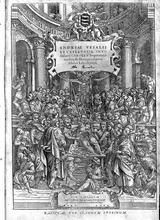
Jan Stephan van Calcar (attributed artist), "Frontispiece," Andreas Vesalius, De humani corporis fabrica libri septem [1555 ed.]. Courtesy of the Wellcome Collection.
At the centre of the celebrated frontispiece of Andreas Vesalius's De humani corporis fabrica (1543) — which heralded modern anatomy — is a half-dissected female body. The real-life model, upon whom Vesalius (1512-1564) demonstrates in a crowded anatomical theatre, was a convicted woman who had staved off a death sentence with a claim of pregnancy. When midwives determined this was not the case, she was executed. Although there are few female bodies in Vesalius's Fabrica (they were difficult to obtain and male anatomy was the standard), anatomists and artists were fascinated by the secrets of woman: the hidden intricacies of her reproductive organs, the mysteries of gestation, and, as the real-life story of Vesalius's subject suggests, her furtive character.
Early modern anatomists and artists mapped the pregnant body in anatomical atlases, and then, in the eighteenth and nineteenth centuries, they modelled it in ivory, wood, leather, textile, terracotta, wax and papier-mâché. Produced by men and women, models embodied classical idealism and scientific rationalism, were objects of education and entertainment, and they travelled across national borders—between France and Japan, Britain and America, the Netherlands and Russia, Italy and Austria. For all of their variety, they shared the quality of virtuality. In fact, Marcel Proust's description of the virtual quality of memory could apply as well to anatomical models: "real but not actual, ideal but not abstract" (3: 888).
Touching and Pointing: Ivory Manikins
For centuries in China, a delicate ivory doll that fit the palm of the hand was used as a communication device between modest female patients who would not speak of their bodies and physicians who were forbidden to examine them. In place of touch and speech, a woman might, from behind a screen, point out her pains on a "doctor's lady." In the nineteenth century, Cantonese carvers produced versions of these reclining, lotus-footed figures as objects of curiosity for Americans and Europeans. They are not dissimilar to European anatomical manikins that were produced mostly in Germany from the seventeenth to the mid-twentieth centuries. Perhaps used to provide rudimentary instruction about pregnancy to young wives, manikins were also decorative collector's items. The arms of some models could be raised over eyes or mouth, allowing the user to lift the abdominal surface to reveal tiny ivory organs and a fetus attached with red umbilical string.
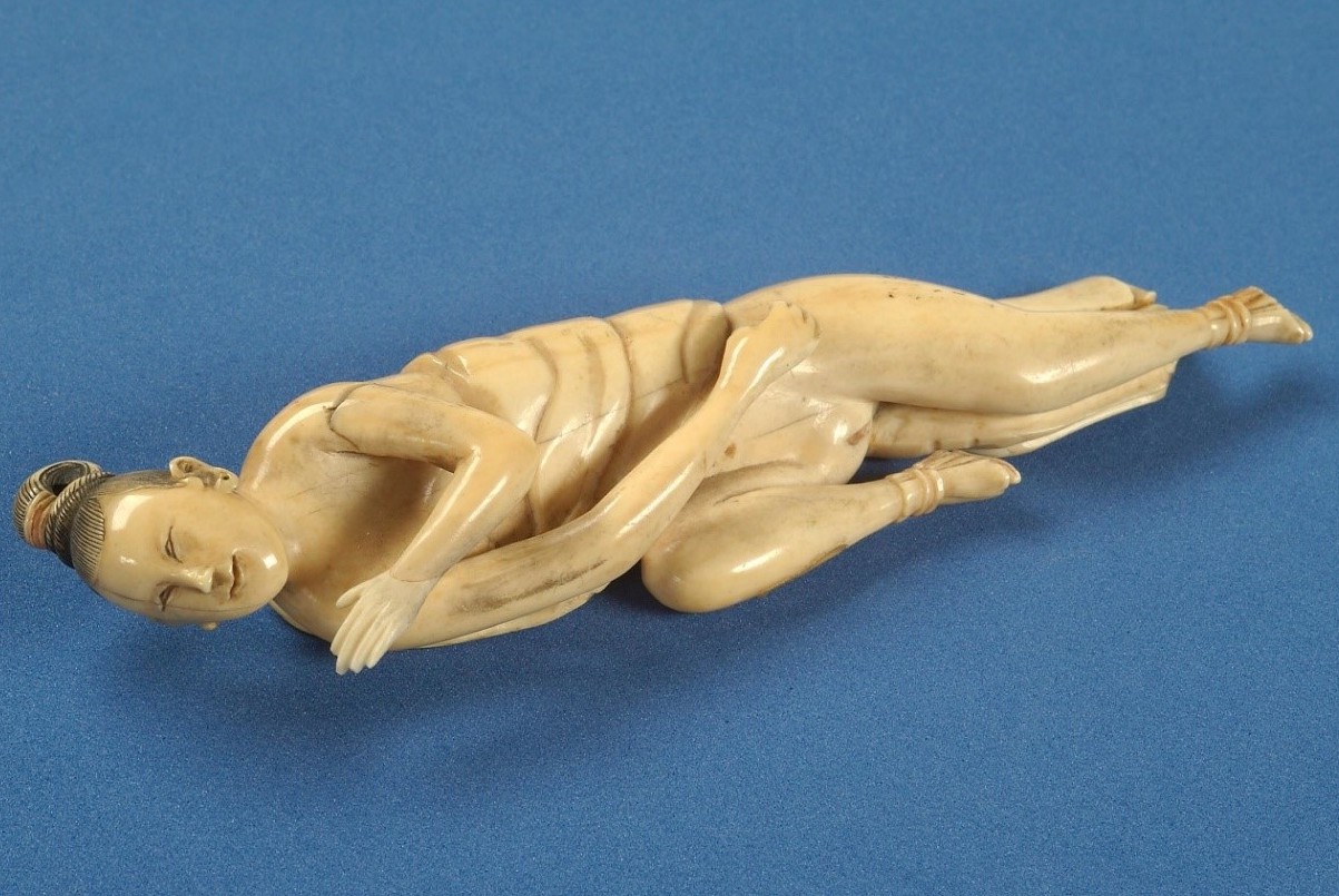
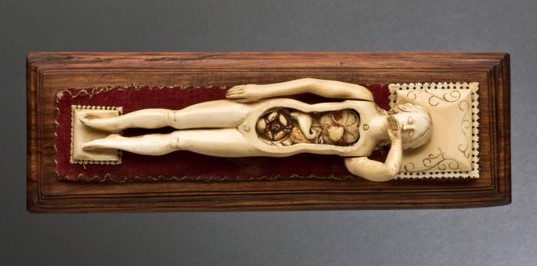
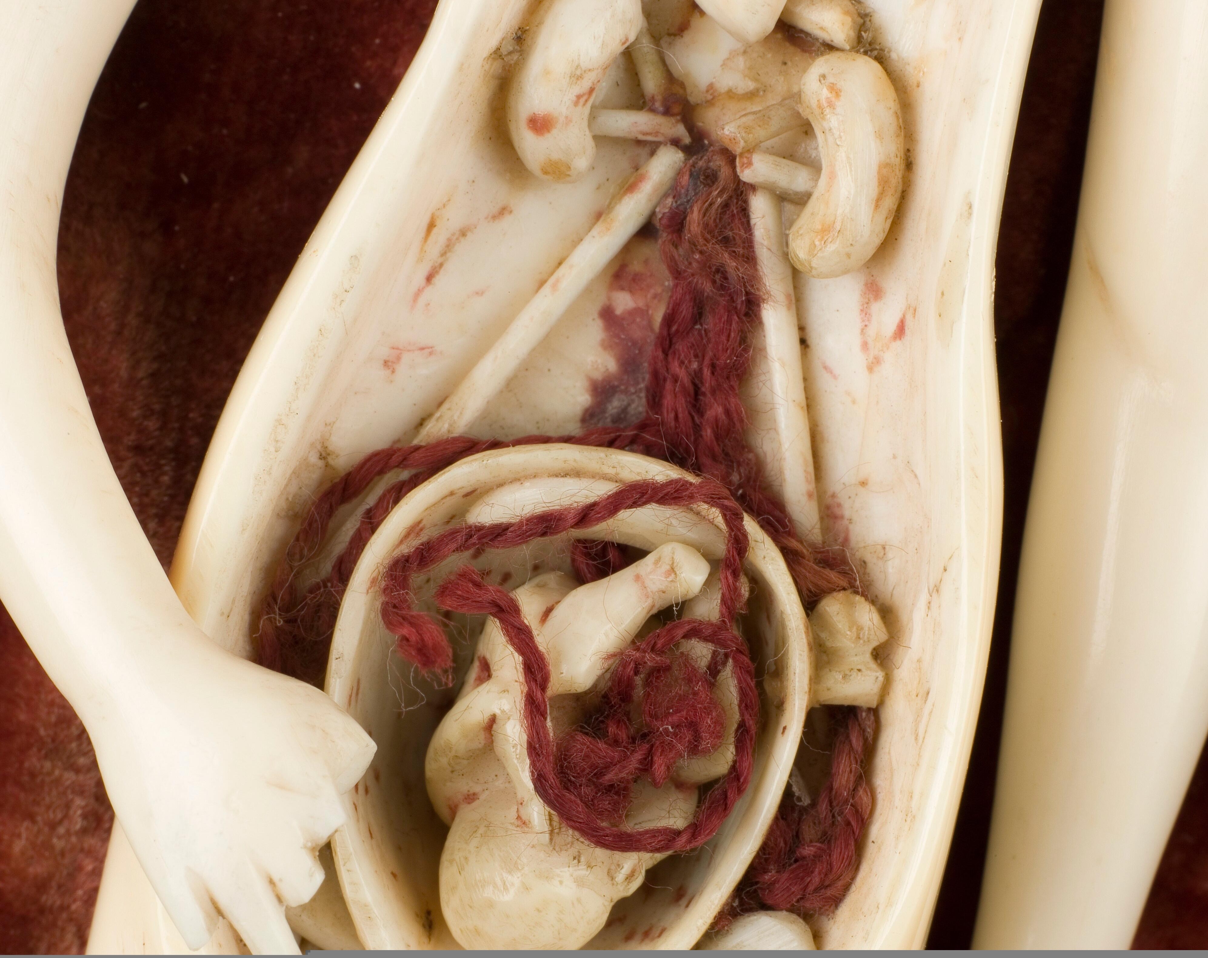
Left: Ivory Chinese diagnostic doll, c. eighteenth-early nineteenth century. Courtesy of the Wellcome Collection. Right two: Ivory anatomical manikins, Europe, possibly Germany, undated, courtesy Science Museum, London (on the 4.0 International (CC BY 4.0) licence).
Vision and Virtuality: Anatomical Venuses
As wax and leather mimicked flesh in ways that ivory did not, while hidden mechanical devices could make more complex, life-sized models seem to breathe with life. In the 1730s, Londoners could view wax anatomical models made by the French surgeon Guillaume Desnoues (d. 1735), a preserved body of a real woman "gone nine Months with Child," and surgeon Abraham Chovet's (1704-1790) model of a pregnant woman, fitted with a mechanism that replicated respiration and circulation. Red liquid moved through glass veins and arteries that connected mother, fetus, and placenta (Daily Journal; Daily Post). When Chovet's model was exhibited at the museum of eccentric inventor and craftsman Benjamin Rackstrow (c. 1707-1772), an advertising pamphlet warned men and women of this virtual vivisection, for she appeared to be a woman "opened when alive," an act that if real would be "of the highest Barbarity and Cruelty" (n.p).
Rackstrow bequeathed his collection to the midwife Catherine Clarke, under whose administration it became a much expanded "anatomical exhibition" in the 1780s. Amongst rooms of curiosities, audiences could see preserved specimens of "diseased wombs," stillborn children, "monstrous births" and wax models of pregnancy. A 1790s catalogue described one of Desnoues's wax models: "Agony" was "beautifully expressed in the Face," while veins could be seen "creeping under the white Skin" and the "minutest Branches" of blood vessels across the "largely distended" womb (10-11).
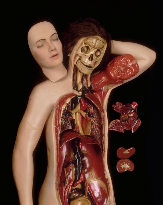
This fusion of idealism and graphic realism, which recalls earlier cultural traditions in which desire and death, beauty and decay, eroticism and spiritualism were not incompatible, also characterized the celebrated Italian wax models known as the anatomical Venuses. Among the more celebrated of these are the "Venerina," or Little Venus, at Bologna and the "Medici Venus" at Florence (see below). Like the ivory manikins, these are "demountable": they can be dissected, from skin and ribcage to lungs, heart, and intestines, and finally, to the uterus with its tiny, curled fetus. The Venuses embody Enlightenment rationality and a prioritizing of "truth-to-nature" — though not at the expense of art, for they celebrate sensory pleasure as much as they commend the senses as conduits of knowledge.
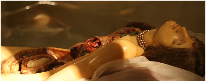
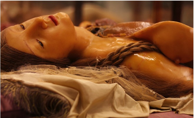
Left: Clemente Susini (artist), La Venerina, 1782; Museo di Palazzo Poggi, Bologna. Right: Clemente Susini and workshop (maker), Anatomical Medici Venus, La Specola, Florence, 1780-82.
The character of wax is complex: the stuff of religious effigies and votives, as well as of Baroque figurines of plague victims; to the touch, it is distinctly earthy, pliable, and sweaty like skin. To the eye, the closed, whole models evoke classical sculpture and biblical narrative, while their open-mouthed expressions, pearls, real hair, and silk beds invite a carnal touch (late in the eighteenth century, locks were fitted on their cases). But the boundary between surface and interior collapses when the models are opened to reveal shiny-moist wax viscera, deeply-coloured in the liver-browns, livid-blues and rust-reds of organs and veins. In 1792, the Florentine Venuses reminded the eighteenth-century painter Élisabeth Vigée-Lebrun of God's divine plan, but the haunting sight of their insides remained superimposed on her vision of living women for some time (1: 237-8). Similarly, when the American novelist Nathaniel Hawthorne visited these models in 1858, he sought relief from their disturbing anatomies by concentrating on "the wholeness and summed-up beauty of woman" (2: 24).
Practical Touching: Birthing Machines
As "Phantoms" or birthing "Machines made in Imitation of real Women," as the Scottish obstetrician William Smellie (1697-1763) described them, invited exploratory touch and trained the hands in methods of delivery. Although Smellie is often credited with inventing the first birthing machine, the highly acclaimed French midwife Angelique de Coudray (1712-1794) got there first in 1758. Her leather and cloth model—with which she travelled all over France—had a flesh-like pliability, boning to give it structure, internal straps that mimicked the birth canal's elastic muscles, and an infant doll with nose, ears and an open mouth that allowed midwives to insert two fingers to practice breach deliveries (Figure 7). Over the years, Coudray developed more elaborate models, including "wet" ones that contained coloured fluids.
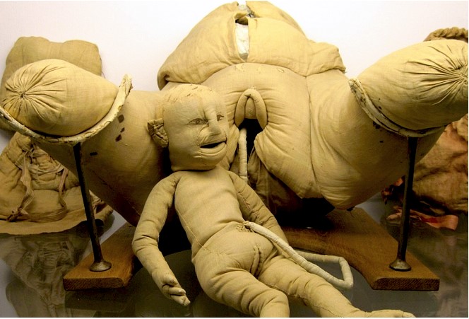
Birthing machine of Madame de Coudray; mid-18th century, Musée Flaubert et d'Histoire de la Médecine à Rouen.
A modern tableau of oil-painted terracotta, Mauro Mazzali's The Obstetrics Class of Giovanni Antonio Galli (2004), imagines the eighteenth-century Bolognese surgeon Galli (1708-1782) demonstrating on a miniature version of an original glass birthing machine (see below). Emphasizing the importance of touch in his teaching, Galli had midwives practice deliveries while blindfolded. In this interpretation of a teaching scene, a midwife physically manipulates a pregnant woman, while Galli maintains a clinical gaze and a professional distance—his hand hovering over the artificial womb—suggesting his membership in a professionalizing fraternity of male obstetricians which increasingly excluded women.
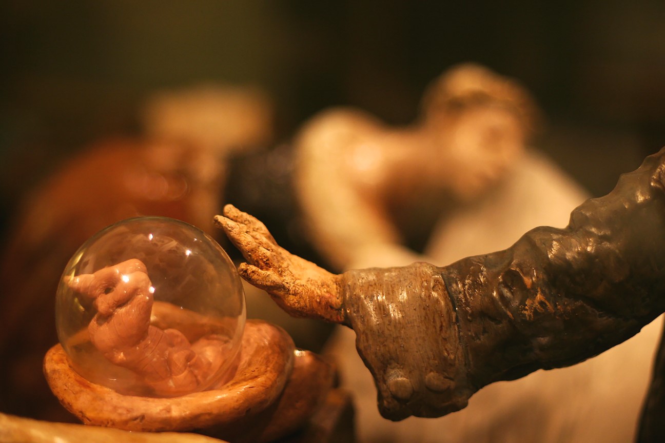
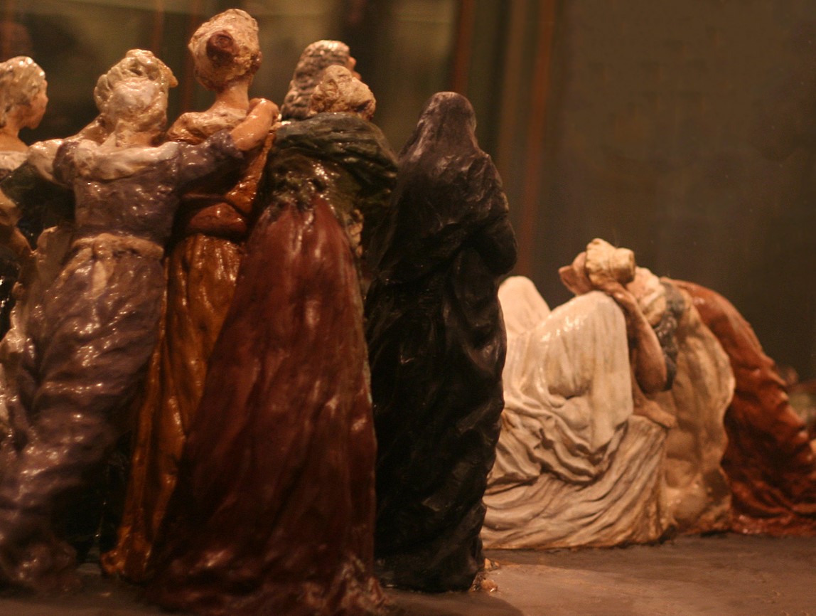
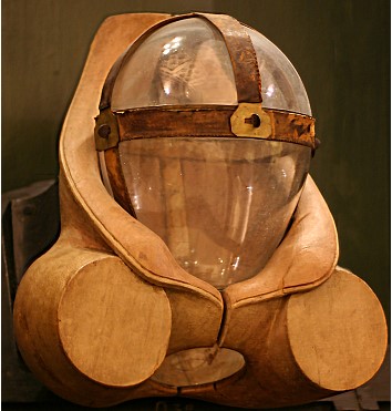
Left two: Mauro Mazzali (artist), The obstetrics class of Giovanni Antonio Galli, 2004, terracotta model; Museo di Palazzo Poggi, Bologna. Right: Giovanni Galli (designer), Antonio Cartolari (creator), Glass birthing machine, mid-18th century; Museo di Palazzo Poggi, Bologna.
The Machine Dismembered
Mechanistic views of the body inform not only the design of birthing machines but also wax and clay models of reproductive organs and fetuses, excised of extraneous parts. In the Bolognese clay model of twins in-utero and in the Florentine ceroplastics below, all signs of personhood are gone and the mother is reduced to a fleshly husk, opened to reveal the secret world of generation.
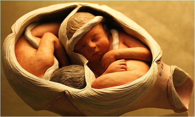
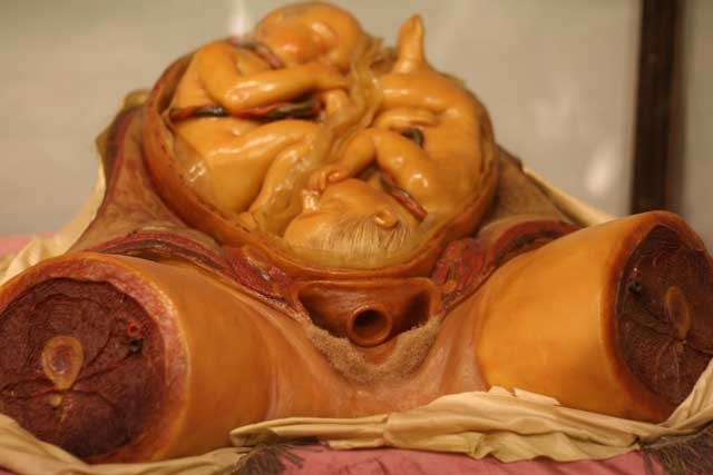
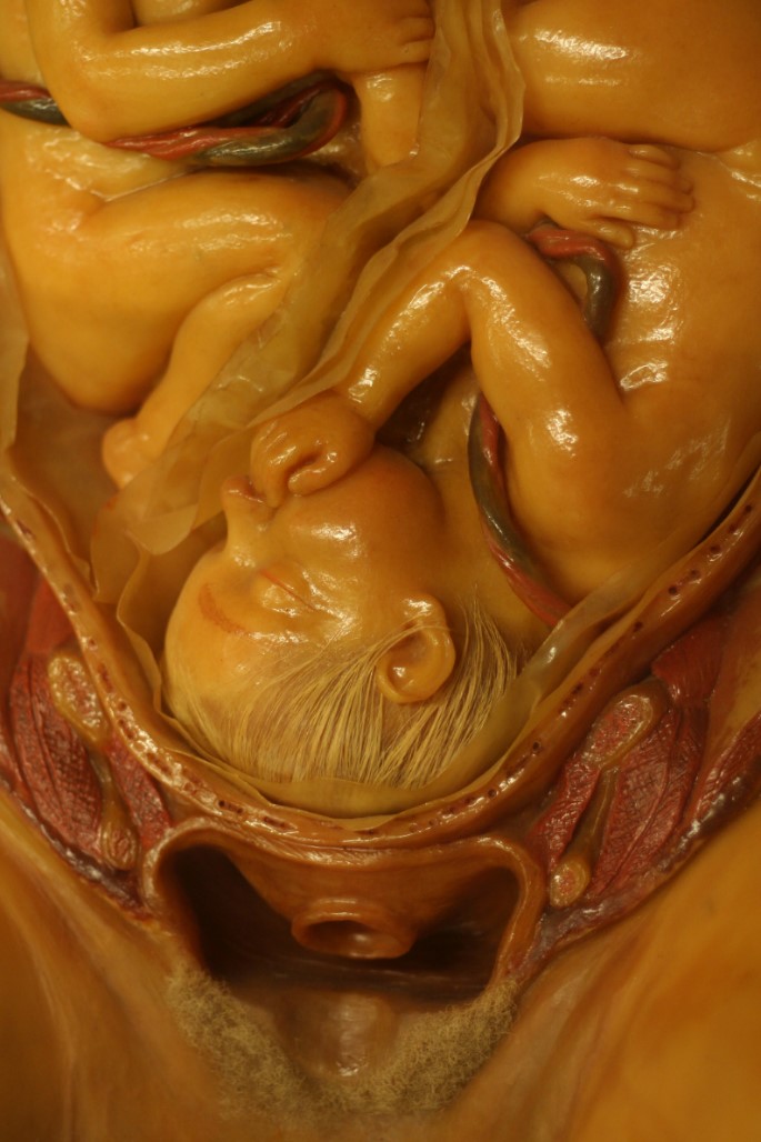
Left: Giovanni Battista Sandri (artist), Giovanni Galli (commissioner), Obstetrical model, polychrome clay, c. mid-eighteenth century, Museo di Palazzo Poggi, Bologna. Middle and right: Clemente Susini and workshop (artists), after William Smellie, Twins in utero, c. 1770–90, La Specola, Florence.
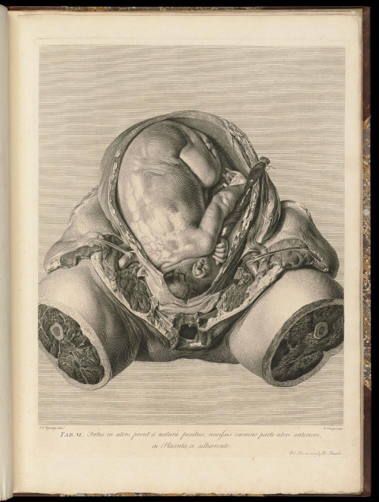
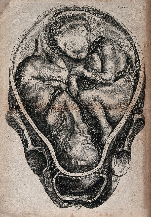
Left: Engraving after William Smellie (anatomist) & Jan van Rymsdyck (artist), "A cross-section of a pregnant uterus containing twins," A set of anatomical tables, 2nd ed. 1761. Right: Jan van Rymsdyck (artist), William Hunter (anatomist), "Fetus in utero." The anatomy of the human gravid uteris, 1774, courtesy of the Wellcome Library.
These latter models are based upon the large-scale naturalistic illustrations of the famous obstetrical atlases of William Smellie and fellow Scottish anatomist William Hunter. Largely drawn by Dutch artist Jan van Rymsdyk, seem to be unmediated images, a "snapshot" of an exact moment of dissection, without artistic flourish. However, such hyper-realistic representation — whether drawing/engraving or model — required careful behind-the-scenes artistry and many numbers of dissections to produce a composite image.
Art and Science; Entertainment and Education
The models above, of uterus and fetus, and of dismembered mother, declare a more obvious allegiance to truth-to-nature than the full-figured, highly aestheticized anatomical Venuses. In the nineteenth century, such differences in illustrative priorities became more pronounced, and the centuries-long alliance between science and art weakened. These changes were reflected in models of pregnancy: there were those, intended for medical audiences, which aspired to mechanical objectivity; then, there were those, intended for popular audiences that sought largely to entertain.
Of the scientific type could be counted the precise papier-mâché obstetrical models produced at anatomist Louis Auzoux's progressive factory, a 'production Utopia', in a Normandy village. The 1850s ovology set below, which includes a cross-section of the vagina and ovaries, as well as twenty-one models showing embryonic development from ovulation to the thirtieth day, instructed students in the new field of embryology (see below). Of the popular, fairground type could be counted Pierre Spitzner's anatomical Venuses, which from mid-century, were exhibited alongside pathological models showing the horrors of syphilis and other 'moral' diseases at the Grand musée anatomique et ethnologique in Paris.
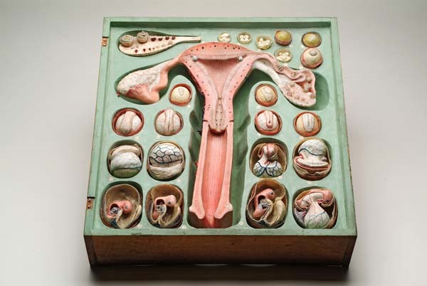
Louis Auzoux, Ovology set, c. 1850; Creative Commons Public Domain Mark 1.0 License.
Among the stars of Spitzner's show were a number of Venuses in glass cases: the Venus Endormie, or Sleeping Venus, the Ceasarian Venus, the 40-piece Demountable Venus (see below), and full-sized figures in the process of delivering or being delivered. Described by writer Italo Calvino as 'the most incredible example of sadist-surrealist fantasy', the Caesarean Venus is a study in strangely violent contrasts and resemblances (29; see below). Bodiless, manicured hands stand out against bloodless, white-gowned, doll-mother whose own hands are absent and whose ankles are bound; then, her oval face mirrors a gaping abdominal cavity of similar shape and size. Calvino reads her expression as 'pain', but it could be read rather as surprise, an expression that might, in turn, reflect those of visitors to Spitzner's exhibition.
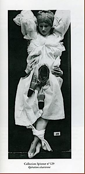
Left: Pierre Spitzner, "Anatomical Venus in 40 parts." Right: "Caesarean Venus," c. mid-19th century, University of Montpellier, France; Creative Commons Public Domain Mark 1.0 Licence.
All of these wonderful models, only a fraction of those produced in the eighteenth and nineteenth centuries, are an intriguing mix of science and art, idealism and realism, fantasy and fact. It stands to reason that they provoke varied responses, from wonder to desire, fear to revulsion. Whatever our own response, these models reflect — and have shaped — how we visualize and understand the workings of this mysterious machine, the body, and its reproductive capabilities.
Bibliography
Daily Journal 3088 (Nov 28, 1730) and 3091 (Dec 2, 1730).
Daily Post 5506 (May 5, 1737).
Calvino, Italo. "The Museum of Wax Monsters," Collection of Sand, trans. Martin McLaughlin, Boston: Mariner, 2002, 26-31.
Hawthorne, Nathaniel. Passages from the French and Italian Note-Books, 2 vols. London: Strahan, 1871.
Proust, Marcel. Remembrance of Things Past, trans. C. K. Scott Moncrieff. 3 vols. London: Penguin, 2016.
Rackstrow, Benjamin. An Explanation of the Figure of Anatomy. London: 1747.
Rackstrow Museum. A Brief Description of Those Curious and Excellent Figures of the Human Anatomy in Wax. London, 1790.
Smellie, William. A Course of Lectures upon Midwifery, […] Perform'd on Different Machines made in Imitation of Real Women and Children. London: 1745.
Vigée-Lebrun, Louise-Élisabeth. Souvenirs, 2 vols. Paris: Charpentier, 1869.
Created 23 September 2022